Blood Pressure: Systolic decreases / Diastolic decreases (peripheral resistance decreases)
Femoral Vein Pressure increases(3x)- Varicosity and Hemorrhoids
Plasma volume increases(7500 ml)
Heart rate ,stroke volume and thus cardiac output increases.(CO=SV*HR)
Hematology Changes-
Red cell mass ,WBC count, ESR increases
Hypercoagulable state
Hemoglobin decreases due to dilution by increased plasma volume-Physiologic anemia
Skin Changes-
Striae Gravidarum(Stretch marks),
.jpg)
Palmar erythema
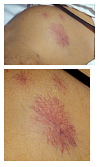
spider anigomas( increased estrogen),
Chadwick sign ,
Linea nigra

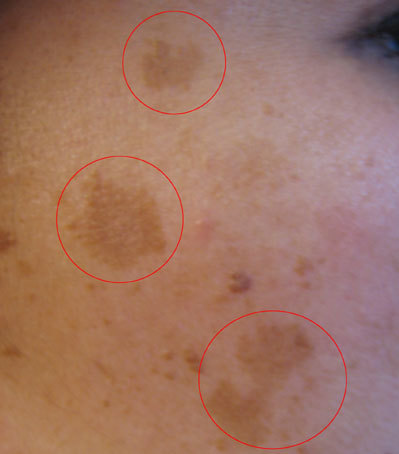
Chloasma(melasma)
Gastrointestinal Changes-
Smooth muscle tone and motility decreases (progesterone)causing constipation , GERD, Cholelithiasis
Lung Changes -
Tidal Volume increases
therefore Minute ventilation increases- causing respiratory alkalosis
Renal Changes-
Kidney size increases, ureter diameter increases(right common), UTI increases
Renal Plasma Flow ,GFR, Creatinine Clearance increases.
BUN,Serum creatinine,Serum uric acid decreases
Glucose increases in urine
Endocrine Changes:
Pitutary size increases( increased blood flow)
Cortisol increases, Thyroid gland size increases, Total T3&T4 increases because there is increase in Thyroid Binding Globulin.

